The animated GIF image is obtained from two frames (more than two for better results), i.e. X-rays pictures of the same subject, but taken from slightly different angles. This way the image does not contain any fictitious information and it can be useful, especially to understand the relative positions of the different elements. This technique has the advantage of not requiring anything new since it is based on already existing tools and software. Of course, it would be better to have an X-rays tool taking the two pictures at the same time, but in most cases a slight rotation of the subject is enough and it allows to spare some money.
(NB: I have no economic interests in this field).
For this video and radiological technique, I humbly suggest the name wigglex, i.e. the word crossing of "wiggle (stereoscopy)" and "X (rays)”.
Wigglex could be useful, especially to understand the relative positions of the different elements, e.g. the position of a foreign body (a bullet, for example) compared to an organ. A standard radiography flattens everything on a 2D picture (radiograph): you cannot understand what is in front and what is behind. For example,the radiograph of a foreign body in front of the liver is the same as a radiograph of a foreign body behind the liver.
In my research, for several X-rays pictures, I’ve been helped by SGP Testing SRL, which I thank, a company dealing (among other things) with radiographs of industrial products for quality assurance.
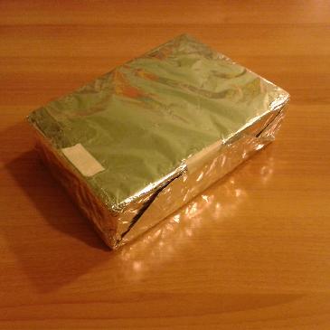
For example, let’s consider this vaguely parallelepipedal object containing some little balls.
This is an approximately frontal image:
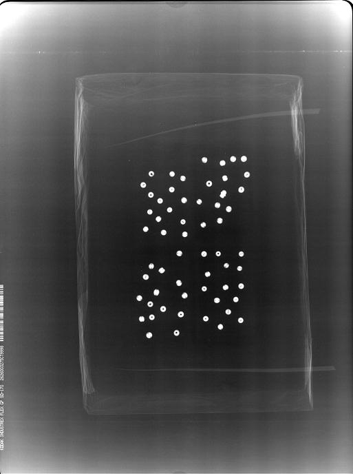
Not even a second approximately frontal image
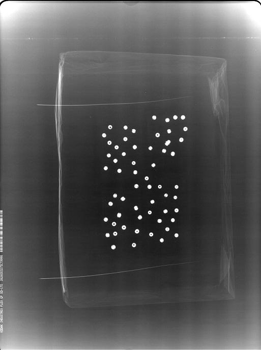
can change the idea that the object contains some little balls in a vaguely rectangular zone.
Let’s have a look now at this radiograph, taken from the long side of the parallelepiped:
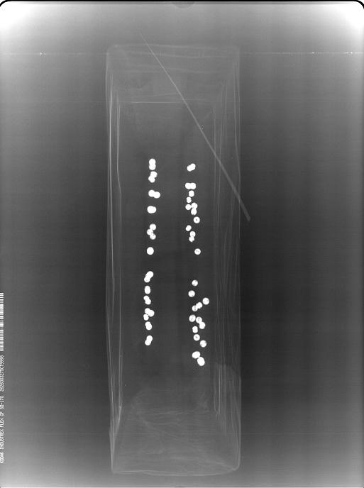
The last radiograph, together with all the previous ones, gives the idea that the little balls are located (approximately) in two parallel rectangles.
This further radiograph, taken in the direction of one of the smaller sides of the parallelepiped,
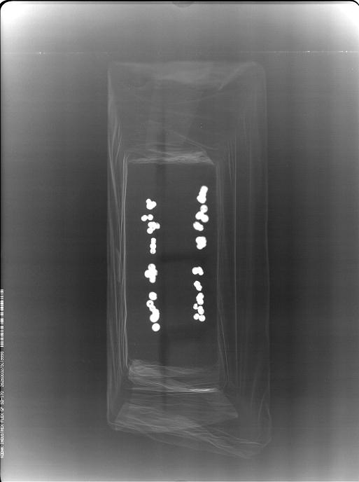
confirms the same idea.
But are the the little balls really arranged this way?
No, they are not!
If you want to know how they are really arranged, click here.